pleural effusion cat ultrasound
Diagnostics will be necessary to confirm the cat has pleural effusion and determine a cause. In some cats infection with mutated coronavirus can lead to blood vessel damage which results in fluid leakage.

Midscapular Thoracentesis Ultrasound Training Model Ultrasound Training Ultrasound Emergency Medicine
Treatment Pleural Effusion in Cats.

. Cats presenting with pleural effusion are nearly always in respiratory distress ranging from an increased respiratory rate and effort to open mouth breathing. Accumulation of fluid in the pleural space. A chest ultrasound to look for the presence of fluid within the pleural cavity.
Feline infectious peritonitis. Measurement of a pleural effusion volume with point-of-care ultrasonography may be a useful tool for intensivists and is an active area of research in critical care 7. Determining the underlying aetiology is.
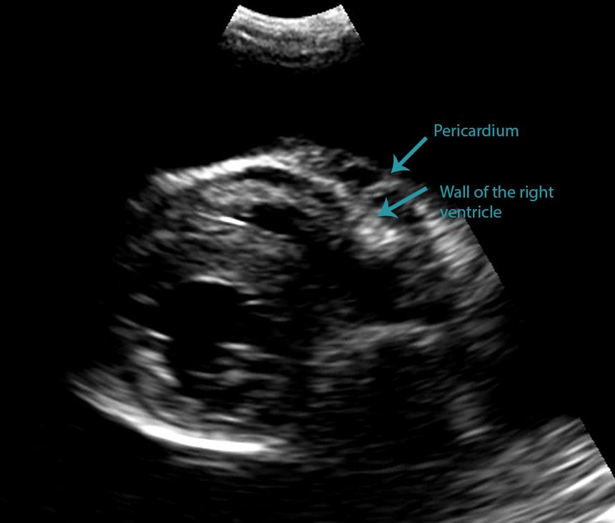
Blood NTproBNP LUS and FCU evaluating left atrial LA size and presence of pericardial effusion PCEFF were performed in all cats. Unlike with a pericardial effusion in the case of accumulation of fluid in the pleural space there is no collapse of the heart walls. Tumors in the lungs or chest wall can lead to pleural effusion.
Ultrasound examination of the heart echocardiogram Laboratory tests. Pleural effusions are characterized on CT by attenuation values between those of water 0 Hounsfield units HU and soft tissue approximately 100 HU typically in the order of 10 to 20 HU. Pleural effusion or pericardial effusion can cause muffled heart sounds.
Vet Radiol Ultrasound 1998. In controlled settings ultrasound may detect constitutive pleural fluid can reliably detect effusions 20 mL in clinical settings and may approach the sensitivity and specificity of computed tomography. When a cat is suffering from pleural effusion the liquid present in the chest cavity prevents the lungs from fully inflating.
Some affected cats may also cough. TFAST a standardized and validated thoracic point-of-care ultrasound examination includes 5 acoustic windows. Sitting or lying in strange positions to ease breathing.
Examination of the effusion included determination of specific gravity using a refractometer Atago Company as well as measurement of the total cell count with the Cell-Dyn 3500 System Abott Laboratories. TFAST Accurate Diagnosis of Pleural and Pericardial Effusion Caudal Vena Cava in Dogs and Cats. Given that most effusions are detected by x-ray which generally cannot distinguish between fluid types the fluid in.
In the latter situations therapeutic intervention must be initiated quickly to prevent respiratory arrest. This syndrome is caused by infection with a mutated form of a feline coronavirus. Abdominal ultrasounds were performed in 70 cats with pleural effusion and revealed concurrent abdominal effusion in 59 of these cats.
This review outlines a practical approach to cases of pleural effusion focusing on early recognition and confirmation of pleural space disease stabilisation of the patient and logical diagnostic investigation. Initial treatments may vary depending on the likelihood of the specific diseases based on your pets physical examination and history. When FIP affects the chest cavity pleural effusion results.
This can be caused by thoracic lymphangiectasia swollen lymph vessels that leak chyle into the pleural space congestive heart failure obstruction of the cranial vena cava the major vein that returns blood to the heart from the front of the body cancer fungal infection feline heartworm. Pleural effusion can have a number of different causes including diseases of the heart lungs or other systemic diseases. The most commonly diagnosed cause of pleural effusion in cats is chylothorax.
A cat with this condition might show some or all of the following signs. Collection of pleural effusion was performed by blind or ultrasound-guided thoracentesis. Ultrasound is widely considered to be more sensitive than radiography to the presence of pleural effusion in man but this has not so far been reported in cats or dogs1 2 3 It is also often used to subjectively monitor fluid volume in cases of chronic effusion.
Pleural effusion can be confirmed with radiography a single DV view if patient permits or thoracic ultrasonography. There is no published method to reliably quantify pleural fluid volume in cats although methods are. In the following article we present two cases concluding with a third case in which both types of effusion can be seen simultaneously.
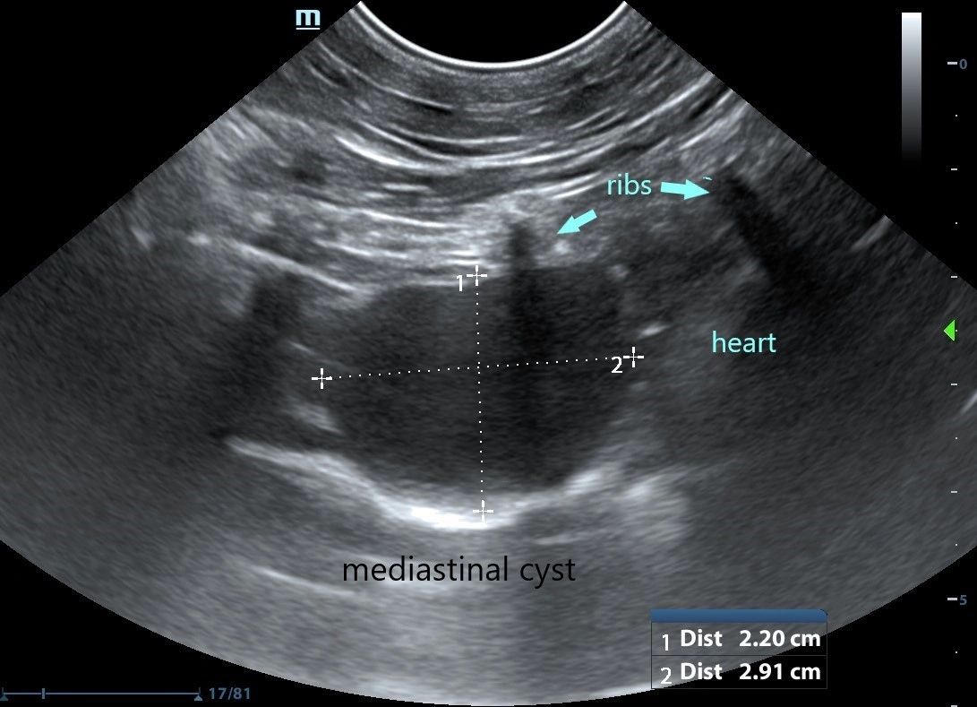
The lack of specificity is mainly due to the limitations of the imaging modality. Cats with pleural effusion often have rapid shallow breathing and pet owners may notice increased respiratory effort. For those who are new to imaging around the heart with ultrasound differentiating a pericardial from a pleural effusion can be tricky particularly when the pleural effusion is circumferential around the heart.
Lung ultrasound findings including pleural effusion PLEFF number of Blines and subpleural abnormalities were noted. Pleural effusion is commonly used as a catch-all term to describe any abnormal accumulation of fluid in the pleural cavity. Diverse disease processes result in sufficient fluid accumulation within the pleural space to cause respiratory compromise.
Four standard effusion types recognized in addition to blood. Pleural effusion is typically. Pleural effusion in cats with pyothorax in.
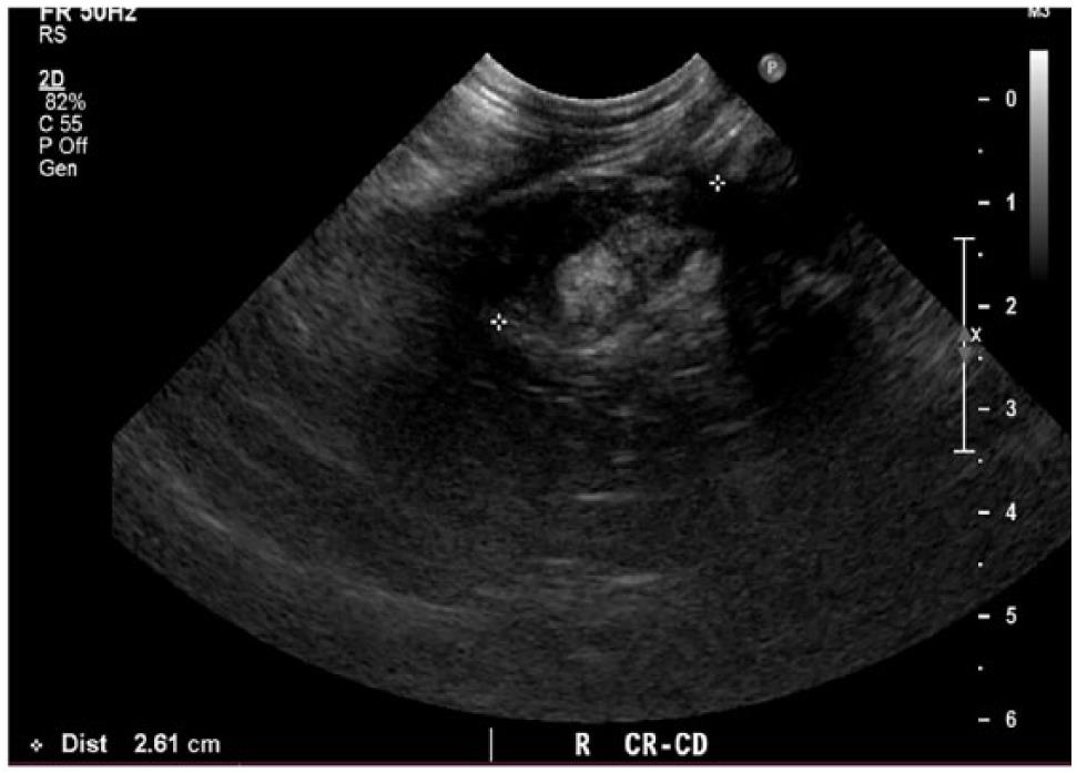
Sagittal ultrasound image of the right upper quadrant shows an anechoic fluid collection above the right hemidiaphragm. Focused Assessment Sonography for Trauma FAST procedure. Medical records were evaluated for final diagnosis.
Major Differential Diagnoses for Pleural Effusion in the Cat. Determining the underlying aetiology is key to appropriate management. This non-invasive and quick test can help the veterinarian evaluate the cat quickly.
This review outlines a practical approach to cases of pleural effusion focusing on early recognition and confirmation of pleural space disease stabilisation of the. Abdominal abnormalities identified on ultrasound included abdominal masses lymphadenopathy hepatic venous congestion hepatomegaly splenomegaly renal enlargement small intestinal wall thickening steatitis and pancreatitis. Diverse disease processes result in sufficient fluid accumulation within the pleural space to cause respiratory compromise.
In the below clip from the Sonoscape S2 you can actually see the separation of the right ventricular free wall from the pericardium in a cat. Rishniw M Weidman J Hornof WJ Hydrothorax secondary to a perinephric pseudocyst in a cat. Screening for effusions can be.
Signs of Pleural Effusion in Cats. Found with right congestive heart failure obstruction to lymphatic drainage by tissue adhesions in pleural space lung lobe torsion neoplasms and abdominal contents herniating. The treatment of pleural effusion ultimately will depend upon the underlying cause.
Bilaterally applied chest tube site and pericardial site views plus diaphragmatico-hepatic view also part of AFAST Vet BLUE. The therapeutic intervention also provides your first diagnostic test. Cats may develop open-mouthed breathing in an effort to increase air flow.

Different Types Of Pleural Effusion On Ultrasound Scan A Exudate B Download Scientific Diagram

Convex Probe Consolidated Lung Surrounded By Hypoechoic Pleural Download Scientific Diagram
Thoracic Ultrasound

Pleural Effusion Radiology Reference Article Radiopaedia Org
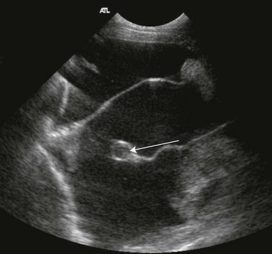
Spontaneous Cholecystopleural Fistula Leading To Biliothorax And Sepsis In A Cat

How To Ultrasound Detection Of Pleural Fluid Case Study Video Youtube

Cardioechography Sonography Student Ultrasound School Ultrasound Physics
Large Secundum Asd With Right Sided Enlargement Sonography Student Heart Function Happy People
Thoracic Ultrasound

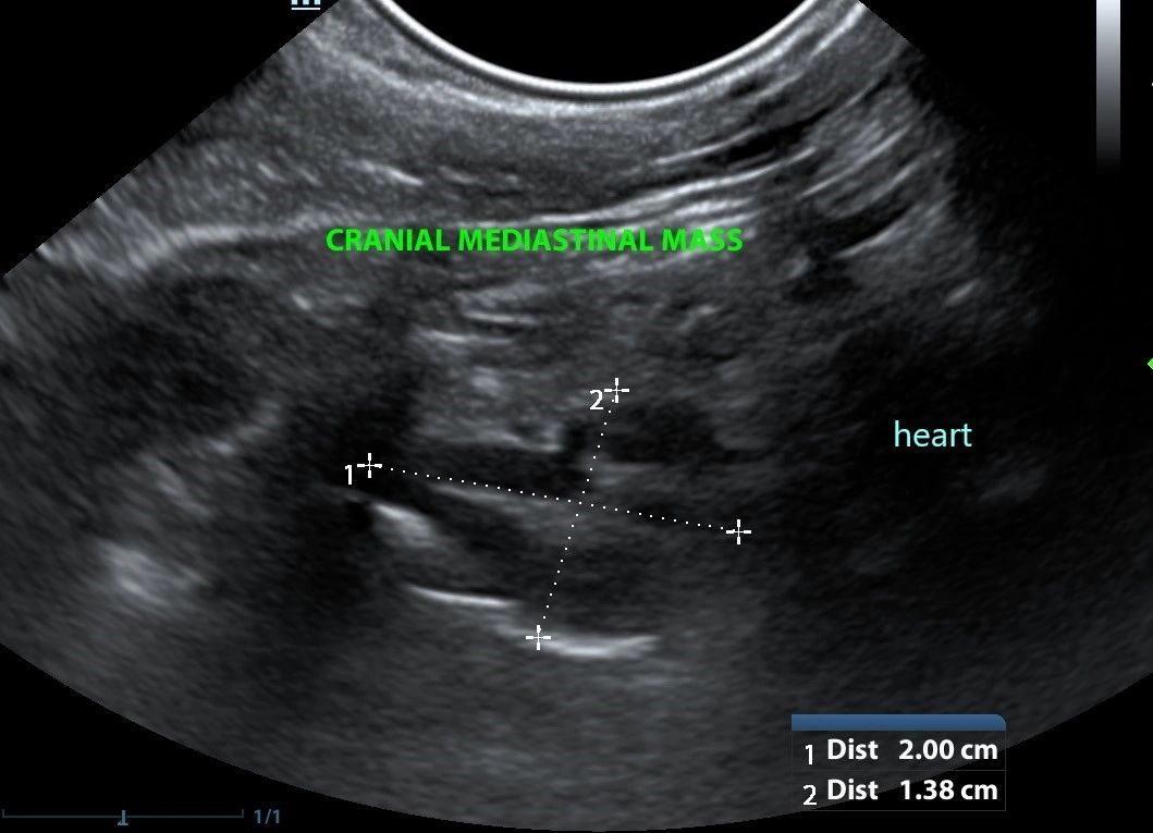
Cat Of Figure 1 Thoracic Ultrasound Revealed A Mild Hypoechoic Download Scientific Diagram

Veterinary Echocardiography Newsletter 1 Effusions Animal Ultrasound Association

Midscapular Thoracentesis Ultrasound Training Model Ultrasound Training Ultrasound Emergency Medicine

How To Ultrasound Detection Of Pleural Fluid Case Study Video Youtube

Different Types Of Pleural Effusion On Ultrasound Scan A Exudate B Download Scientific Diagram

Differentiating Pericardial From Pleural Effusion Animal Ultrasound Association

Pdf Thoracic Ultrasound A Method For The Work Up In Dogs And Cats With Acute Dyspnea Semantic Scholar
Pleura Veterian Key

Differentiating Pericardial From Pleural Effusion Animal Ultrasound Association
Veterinary Echocardiography Newsletter 1 Effusions Animal Ultrasound Association